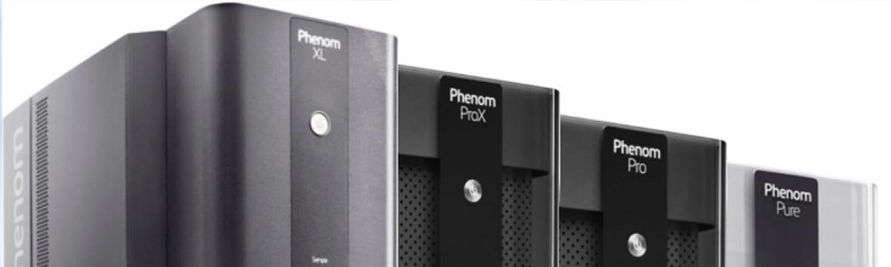
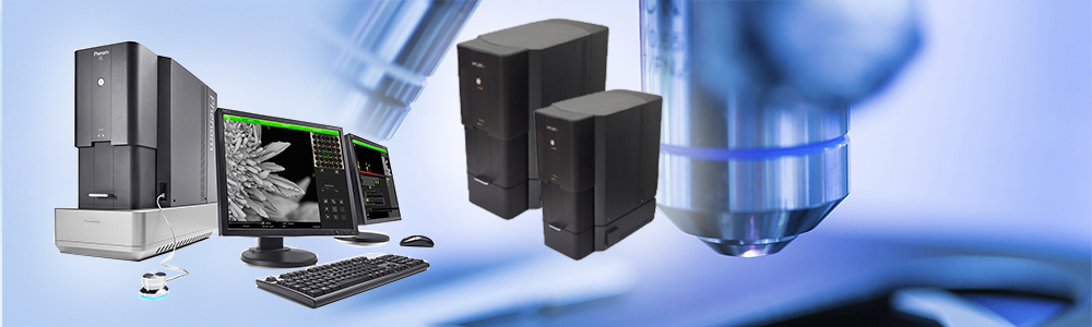
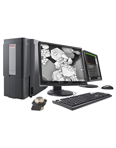
logo

Hotline: +8424 62967539  |  ADD: RM 630, 6th Floor, CIC Tower, No. 2 Nguyen Thi Due Str., Cau Giay, Ha Noi, Vietnam.

sl33
sl44
upt_animation_column_failure
cimps-qe-ipce_wb

Phenom Pharos

Phenom Pharos
phenom-pharos - ảnh nhỏ  1

Phenom Pharos

The Thermo Scientific Phenom Pharos is a desktop SEM with an FEG source that makes crisp, high-brightness images and the benefits of an FEG source accessible to everyone. It is also easy to operate, from the initial installation to the actual usage, thanks to its intuitive and compact design.

The advanced hardware design and detectors enable a fast time to image and easy, foolproof handling.

Key Benefits

High-brightness images

The Phenom Pharos Desktop SEM is designed in such a way that the power of an FEG source, like crisp, high-brightness images, can be accessed by all. 

Optional secondary electron detector

The SED collects low-energy electrons from the top surface layer of the sample. It is therefore the perfect choice to reveal detailed sample surface information. 

High diversity in applications

You can gain more insights with the Phenom Pharos Desktop SEM within specific SEM applications by using a broad range of sample holders, such as metallurgical, temperature controlled, electrical feed-through, and many 3rd party sample holders. 

1606863092614

Specifications

Light optical magnification
  • 20–134x
Electron optical magnification range
  • 200–1,000,000x
Resolution
  • 2.5 nm (SE), 4 nm (BSE) at 15 kV
  • 10 nm (SE) at 3 kV
Digital zoom
  • Max. 12x
Light optical navigation camera
  • Color
Acceleration voltages
  • Default: 5 kV, 10 kV and 15 kV
  • Advanced mode: adjustable range between 2 kV and 15 kV
Vacuum modes
  • High vacuum mode
  • Medium vacuum mode
  • Integrated charge reduction mode (low vacuum mode)
Detector
  • Back scattered electron detector (standard)
  • Energy-dispersive X-ray spectroscopy (EDS) detetor (optional)
  • Secondary electron detector (optional)
Sample size
  • Up to 25 mm diameter (32 mm optional)
Sample height
  • Up to 35 mm (100 mm optional)

Media Gallery

Solar cell imaged with Phenom Pharos Desktop SEM.
Solar cell imaged with the Phenom Pharos Desktop SEM.
SEM image acquired with the Phenom Pharos Desktop SEM.
Gold particles imaged with Phenom Desktop SEM.
Gold particles imaged on a Phenom Desktop SEM.
Red blood cells imaged with Phenom Pharos Desktop SEM.
Red blood cells imaged with the Phenom Pharos Desktop SEM.
Solar cell imaged with Phenom Pharos Desktop SEM.
Solar cell imaged with the Phenom Pharos Desktop SEM.
SEM image acquired with the Phenom Pharos Desktop SEM.

Applications

Process Control_Thumb_274x180_144DPI

Process Control
 

Modern industry demands high throughput with superior quality, a balance that is maintained through robust process control. SEM and TEM tools with dedicated automation software provide rapid, multi-scale information for process monitoring and improvement.


Quality Control_Thumb_274x180_144DPI

Quality Control
 

Quality control and assurance are essential in modern industry. We offer a range of EM and spectroscopy tools for multi-scale and multi-modal analysis of defects, allowing you to make reliable and informed decisions for process control and improvement.


Fundamental Materials Research_R&D_Thumb_274x180_144DPI

Fundamental Materials Research

Novel materials are investigated at increasingly smaller scales for maximum control of their physical and chemical properties. Electron microscopy provides researchers with key insight into a wide variety of material characteristics at the micro- to nano-scale.


SEARCH
SUPPORT ONLINE
icon  +8424 62967539
PARTNER LOGO
8
3
9
zahner_logo_4c_crimson
logo

GSEM., JSC

Tel: +8424 62967539; Email: sales@gsemvn.com

Head Office: RM 630, 6th Floor, CIC Tower, 1 Nguyen Thi Due Str., Cau Giay Dist., Hanoi, Vietnam.

Registered Address: No. 18 Alley 313/6 Lane 313 Linh Nam, Hoang Mai, Hanoi